Structure of Bacteria with Respiration, Reproduction, and Growth
Structure of Bacteria
 All bacteria have cell membrane, cytoplasm, ribosome and chromatin bodies. Most of the bacteria have cell wall. It gives shape to the bacterial cell. Some bacteria have specific structures like capsule, slime, flagella, pill, fimbriae and granules.
[wp_ad_camp_1]
Size
The size and shape vary in different bacteria. Their size is from 0.1 to 600µm over a single dimension.
- The smallest bacteria like some members of Mycoplasma are loo to 200 nm in diameter. Its size is equal is equal to the size of the largest viruses (pox viruses).
- Escherichia coli (a bacillus) has average size from 1.110 1 .5 pm wide and 2.0 the 6.0 pm long.
- Some spirochetes sometimes reach 500 pm in length.
- Staphylococci and streptococci are 0.75 to 1.25 pm in diameter.
- Recently a huge bacterium has been discovered form the intestine of sturgeon fish, Acanthurus nigrofuscus. This bacteria Epulopiscium fishiesoni grows 600/80 pm. It is little small than a printed hyphen. It is now clear that some bacteria are much larger than the average eukaryotic cell.
Shapes of Bacteria
Bacteria are classified in to three categories on the basis of their shapes. These shapes are cocci, bacilli and spiral. Most of the bacteria have constant shapes. Some bacterial cells are pleomorphic and exist in different shapes.
Cocci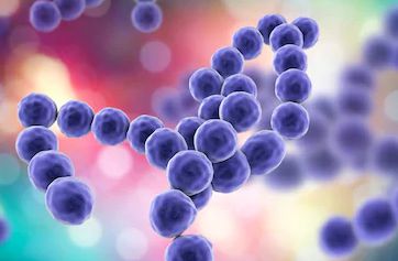
Cocci are spherical or oval bacteria. They have different arrangements. These arrangements are based on their plane of division.
[wp_ad_camp_2]
- Diplococcus: In this case division is in one plane and cocci occur in pairs.
- Strptococcus: In this case, division also occurs in one plane but the cocci forms long chain of cells.
- Tetrad: When division of cells occurs in two planes, it will produce a tetrad arrangement. A tetrad is a square of 4 cocci.
- Sarcina: When division occurs in three planes, it will produce a sarcina arrangement. Sarcina is a cube of 8 cocci.
- Staphylococcus: When division occurs in random planes, it will produce a staphylococcus arrangement. In this case, the cocci are arranged irregularly like clusters of grapes.
Examples of cocci are Diplococcus pneumonia, Staphylococcus aureus.
Bacilli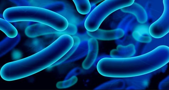
Bacilli are rod shaped bacteria. Bacillus is a single cell of bacteria.
There are following arrangements of bacilli.
- Streptobacillus: Streptobacillus is chain of bacilli.
- Diplobacilli: When bacilli occur in pairs, then the arrangement is called diplobacilli.
Examples of bacilli are Escherichia coli, Bacillus subtilis, Pseudomonas.
Spiral
The spiral shaped bacteria are spirally coiled. There are following forms of spirals:
[wp_ad_camp_3]
- Vibrio: It is a curved or comma-shaped spiral.
- Spirillum: It is a thick rigid spiral.
- Spirochete: It is thin, flexible spiral.
Examples of spiral shaped bacteria are Vibrio, Hyphomicrobium.
Bacterial Cell Structure
Flagella and their Function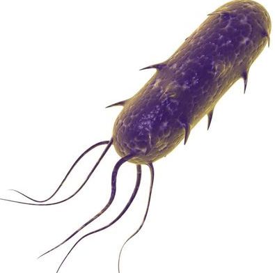
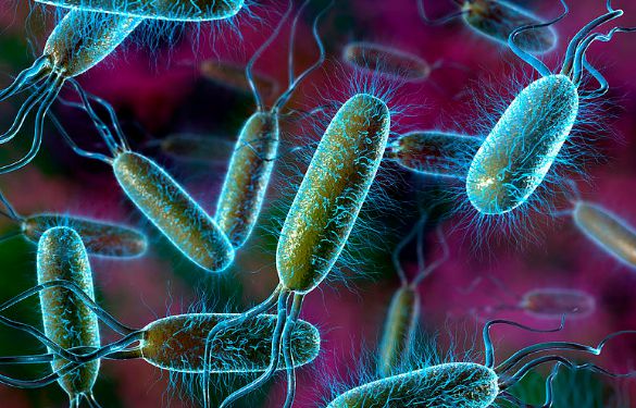
These are extremely thin hair like appendages. They originate from basal body and come out through cell wall. The basal bodies are present just beneath the cell membrane ¡n the cytoplasm. Flagella are made up of a protein flagellin. Most of the bacilli and spiral shaped bacteria have flagella. Cocci rarely have flagella. Bacteria are classified into different taxonomic groups on the basis of presence of flagella, pattern of attachment of flagella and the number of flagella.
- Atrichous: In this case, bacteria are without flagella.
- Monotrichous: In this case, single polar flagellum is present.
- Lophotrichous: In this case tuft of flagella is present only at one pole of bacteria
- Ampitrichous: In this case tuft of flagella is present at each of the two poles of bacteria.
- Peritrichous: In this case flagella surround the whole cell.
The primary function of flagella is locomotion. Bacteria can detect a chemical signals with the help of flagella and move in its response. Such type of behavior is called Chemotaxis.
[wp_ad_camp_4]
Pili and their Functions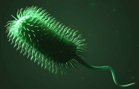
These are hollow, non-helical and filamentous appendages. Pili are smaller than the flagella. They are not involved in locomotion. True pill are present only in gram-positive bacteria. They are made up of special proteins called pilin. They are primarily involved conjugation. Conjugation is a mating process (reproductive). Some pili are used for the attachment of bacteria with various surfaces.
The Cell envelope: The outer wrapping of bacteria
Bacteria have different types of outer wall and surfaces. The complexes of layers, external to the cell protoplasm are collectively called cell envelope. Cell envelop includes capsule, slime and cell wall.
Capsule
Bacteria produce capsule. It ¡s made up of repeating units of polysaccharides or proteins or both. The capsule is tightly bound with the cell wall. It has thick and gummy nature. It makes the encapsulated bacteria sticky.
Slime
Some bacteria are covered with loose, soluble shield of macromolecules. These are called slime. Slime provides greater pathogenicity (power for causing disease) to bacteria. It protects the bacteria against Phagocytosis.
[wp_ad_camp_5]
Cell wall
Cell wall is present beneath the extra cellular substances. It is present outside the cytoplasmic membranes. Cell wall is rigid structure. It determines the shape of the bacteria. Cell wall also protects the cells from the osmotic lysis. Cell wall is absent in Mycoplasma.
Gram staining technique of cell wall
This technique was discovered by Christian Gram. Different bacteria show different response to gram staining procedure. So bacteria are divided into two groups on the basis of this response:
- Gram positive bacteria: They are stained purple with this staining technique. They forms CV- I complex (Crystal violet complex 1) and retain the primary dye colour.
- Gram Negative bacteria: They are stained pink with this staining technique. They retain secondary dye colour.
There are many structural differences between two groups. They are stained differently due to these structural differences.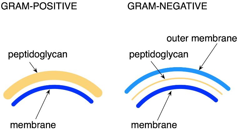
Composition of cell wall
The cell wall of most bacteria has a unique macromolecule called peptidoglycan. Its amount is different in different types of bacteria. It is composed of long framework of glycan chains (glucose). These glycan chains are linked with peptide fragments. The intact cell wall also contains some molecules of sugar, teichoic acid, lipoproteins and Lipopolysaccharides. These molecules are linked to peptidoglycan.
[wp_ad_camp_1]
The cell wall structure present in gram-positive and gram- negative bacteria is absent in some bacteria. Some bacteria have no cell wall at all. The cell wall of Archaeobacteria is different from the Eubacteria. Archaeobacteria do not contain peptidoglycan. Their cell wall is composed of proteins, glycoprotein and polysaccharides.
Cell Membrane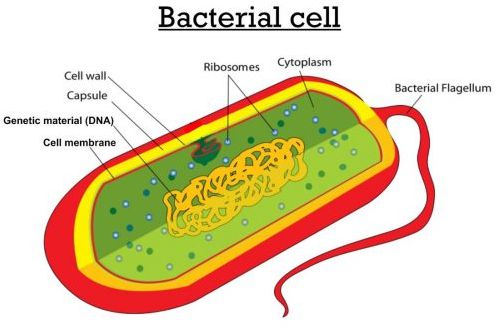
Cell membrane or plasma membrane is present beneath the cell wall. It is very thin and flexible. It completely surrounds the cytoplasm Plasma membrane is very delicate in nature. Any damage to it results in death of the organisms. The bacterial cell membrane is different from the eukaryotic membrane. It lacks sterols like cholesterol.
Functions of cell membrane
Cell membrane regulates the transport of proteins, nutrients, sugar, electrolytes and other metabolites. The plasma membrane also contains enzymes for respiratory metabolism.
Cytoplasmic Matrix
The cytoplasmic matrix is a substance present between the plasma membrane and the nucleoid. The membranous bound organelles and cytoskeleton (microtubules) are absent in the prokaryotic cytoplasm. It has gel like structure. Small molecules can move through it rapidly. The plasma membrane and everything present in it is called protoplast. Thus the cytoplasmic matrix is a major part of the cytoplasm. Other structures like chromatin/nuclear body, ribosomes, mesosomes, granule and nucleoid are present in this matrix.
[wp_ad_camp_2]
Nucleoid
Discrete chromosome and nuclear membrane is absent in bacterial cell. The nuclear material of DNA is present near the center of the cell. This nuclear material is composed of single, circular and double stranded DNA molecule. The nuclear material aggregates to form irregular shaped dense body called nucleoid. This nucleoid is also called nuclear body, chromatin body or nuclear region. It has very long molecule of DNA. This DNA is tightly folded and fit inside the cell components. Bacteria has single chromosome. So bacteria are haploid. Nucleoid is visible in the light microscope after staining with Feulgen stain. Escherichia coli closed circle chromosome measures approximately 14000 pm.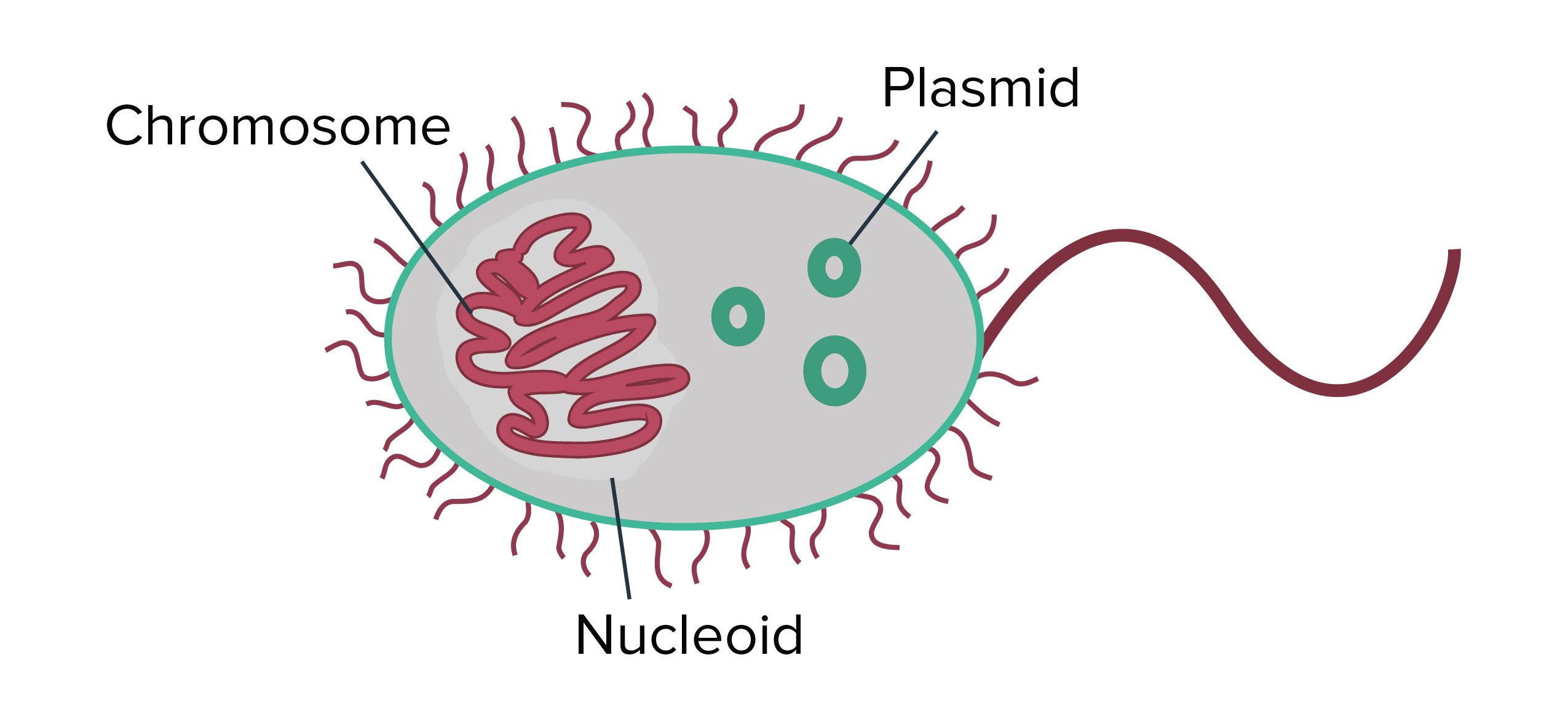
Plasmid
Many bacteria contain plasmids in addition to chromosomes. Plasmids are circular, double stranded DNA molecules. They are self-replicating bodies. Plasmids are not essential for the bacterial growth and metabolism. They contain drug and heavy metals resistant genes. Disease and insect resistant genes are also present on them. Plasmids are uses as vector in modern genetic engineering.
Ribosomes
Ribosomes are composed of RNA and protein. Some ribosomes are also loosely attached with the plasma membrane. They are protein factories. There are thousands of ribosomes in each healthy growing cell. The ribosome of bacteria (70S) is smaller than the ribosomes of eukaryotes (80S).
[wp_ad_camp_3]
Mesosomes
The cell membrane invaginates to form mesosomes. Mesosomes are in the form of vesicles, tubules or lamellae. They perform following functions:
- Mesosomes are involved in DNA replication and cell division.
- Some mesosomes are also involved in the export of exocellular enzymes.
- Respiratory enzymes are also present on the mesosomes.
Granules and Storage bodies
Bacteria live in an environment where nutrients are in short supply. The bacteria try to store extra nutrients when possible. These may be glycogen, sulphur, fat and phosphate. The cell also contains waste material. This waste material is excreted later on. Common waste materials are alcohol, lactic acid and acetic acid.
Spores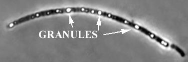
Certain species of bacteria produces spores. There are two types of spores:
- Exospores: These are produced outside the vegetative cells.
- Endospores: These are present within the vegetative cells.
Spores are metabolically dormant (inactive) bodies. They are produced at later stage of growth. Spore are resistant to adverse environmental conditions like light, high temperature, desiccation (dehydration), pH and chemical agents. They grow under favorable conditions and form new vegetative cells.
[wp_ad_camp_4]
Cysts
Cysts are dormant, thick- walled and desiccating resistant structure. They develop during reproduction of vegetative cells. Such cells germinate only under suitable conditions. They are not heat resistant structures.
Respiration in Bacteria
Bacteria are divided into different groups on the basis of their mode of respirations:
- Aerobic bacteria: These bacteria require free oxygen for their respiration. They can grow only in the presence of oxygen, e.g. Peudomonas.
- Anaerobic bacteria: They do not require free oxygen. They can grow in the absence of oxygen, e.g. Spirocheta
- Facultative bacteria: They can grow in presence or absence of oxygen, e.g. E.coIi
- Microaerophilic bacteria: These bacteria require a low concentration of oxygen for growth, e.g. Campylobacteria.
Growth and Reproduction
The increase in number of cells of bacteria is called bacterial growth. Bacteria increase in number by asexual reproduction called binary fission. The parent cell enlarges during binary fission. Its chromosome duplicates and nuclear material is equally distributed in the cell. Its plasma membrane pinches inward at the center of the cell and divides the cell in to two. These divisions are repeated after fix intervals and new daughter cells are formed. The interval of time until the completion of the next division is known as generation time. After the completion of each division, the daughter bacteria grow and develop their unique features. So the population of bacteria is increased.
Phases of growth
There are four distinct phases of growth in the bacterial growth curve:
- Lag Phase: It is phase of no growth. Bacteria prepare themselves for division.
- Log phase: It is phase of rapid growth. Bacteria divide at exponential rate.
- Stationary phase: Bacterial death rate becomes equal to bacterial rate of reproduction and multiplication.
- Death / Delcine: Bacteria start dying. Here the death rate is more than the reproduction rate.
Conjugation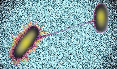
Traditional sexual reproduction (gamete formation) and mitosis is absent in bacteria. Some bacteria transfer their genetic material form a donor bacterium to a recipient bacterium during the process of conjugation. Some conjugating bacteria have specialized sex pili. These pili transfer genetic material. Conjugation produces new genetic combinations. These combinations help bacteria to survive in different conditions.


some truly quality content on this website , bookmarked.
Thank You!