There are two types of microscopes i.e. light microscope (LM) and Electron Microscope (EM) are used in microscopy. the Major Difference between Light Microscope and Electron Microscope is that light microscopes can see reasonable detail with true colors and are good for small organisms, invertebrates, and whole-cell while electron microscopes can magnify highly detailed images and even 3D surfaces and can see organelles of cells, bacteria, and even viruses.
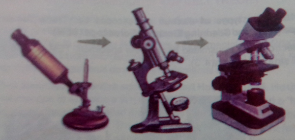
It works by passing visible light through a specimen. It uses two glass lenses. One lens produces an enlarged image of the specimen and the second lens magnifies the image and projects it into the viewer’s eye or onto photographic film. A photograph taken through a microscope is called a micrograph.
Light microscopes can magnify objects only about 1500 times without causing blurriness i.e. its magnification is 1500X. Its resolving power is 0.2 micrometer (µm). And 1µm= 1/1000mm. In other words, the LM cannot resolve objects smaller than 0.2µm. it is about the size of the smallest bacterium. The image of a bacterium can be magnified many times, but light microscopes cannot show the details of the internal structure.
It is the most advanced form of the microscope. In EM, objects and lenses are placed in a vacuum chamber and a beam of the electron is passed through the object. Electrons pass through or reflected from objects and make images. Electromagnetic lenses enlarge and focus the image onto a screen or photographic film.
EM has much higher resolving power than LM. The most modern EM can distinguish objects as small as 0.2 nanometers (nm) and 1 nm= 1/1000, 000 mm. It is a thousand-fold improvement over LM. EM can magnify objects about 250,000 times. Under special conditions, EM can detect individual atoms. Cells, organelles, and even molecules like DNA and protein are much larger than single atoms.
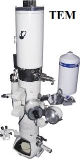
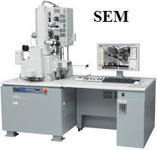
Biologists use two types of electron microscopes i.e. Transmission Electron Microscope (TEM) and Scanning Electron Microscope (SEM).
In TEM, electrons are transmitted through the specimen. TEM is used to study the internal cell structure.
In SEM, electrons are reflected from the metal-coated surfaces. SEM is used to Study the structures of cell surfaces.
When it comes to creativity, you might ask yourself whether or not you can feel…
In the dynamic landscape of India, where tradition and technology frequently intersect, the domain of…
In this article, we will provide a comprehensive explanation of IMDb's basic information, simple usage…
Boostcraft.net is your one-stop destination for all your WoW retail boost needs. Our team of…
California's logistics and warehousing industry has experienced tremendous growth over the years. With the increasing…
In recent years, the food and beverage industry has witnessed a significant shift towards digitization,…